HistoLab
The HistoLab facility provides comprehensive support for tissue processing and advanced imaging.


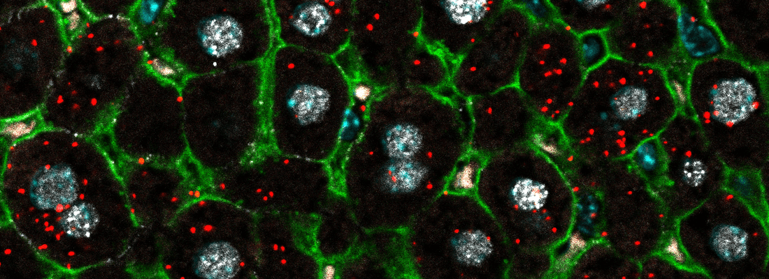
The HistoLab histology facility provides comprehensive support for tissue processing and advanced imaging. We offer conventional histology, immunohistochemistry (IHC), RNAscope, tissue clearing and 3D imaging, as well as assistance with slide preparation for metabolomics and spatial transcriptomics. In addition, we provide guidance with image analysis.
To ensure efficiency and reproducibility, many of our procedures are supported by automated equipment.
The price list for HistoLab's services for external users is available to download here.
Our goal is to make histology accessible, reliable, and innovative. We aim to support researchers at every stage—from project planning to data analysis—by offering state-of-the-art equipment, specialized expertise, and personalized guidance.
We are pleased to welcome all users—regardless of their level of histology experience—from CBMR, the Institute of Neuroscience, BMI, as well as researchers from other institutes, faculties, universities, and hospitals.
For new users
If you are new to the facility, you must first receive an introduction to the histology laboratory and the equipment you wish to use.
- For paraffin embedding, microtome training, or routine staining in the histology lab, please contact Nataliia Tiutcheva.
- For training on microscopes or the cryostat, or for information regarding protocols, antibodies, or probes, please contact Oksana Dmytriyeva. The type of introduction will depend on your previous microscopy experience.
Project support
If you require assistance with your project—including experimental planning, conducting experiments, data analysis, or manuscript preparation—please send a short project description to Oksana Dmytriyeva.
- Guidance for project planning, experiment design, and workflow optimization
- Access to specialized equipment enabling automation of histological procedures
- Technical assistance with tissue processing, embedding, sectioning, staining, and imaging
- Training modules in histological techniques and equipment handling
- Support with advanced methods such as IHC, RNAscope, tissue clearing, and 3D imaging
- Assistance with image acquisition, data analysis, and interpretation
- Embedding station TES99
- Tissue processor Excelsior AS
- Microtome Epredia HM 355S
- Cryostat Leica 3050S
- BOND RX Fully Automated Research Stainer
- HybEZ II Oven for RNAscope
- Zeiss AXIO Imager.Z2 Fluorescence Motorized LED Microscope Microscope
- Zeiss AXIO Observer Z1 Inverted Fluorescence Microscope